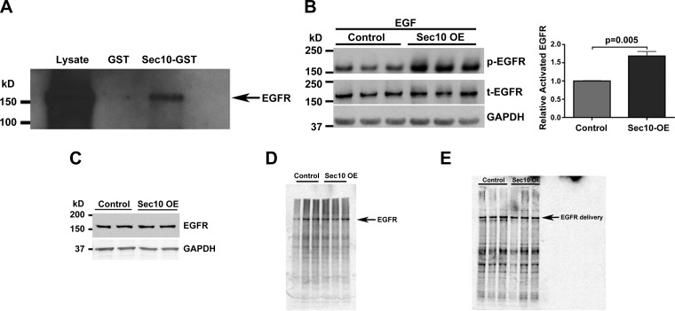
Fig. 1.
Sec10 biochemically interacts with epidermal growth factor receptor (EGFR), and Sec10 overexpression results in more potent EGFR phosphorylation in response to EGF. A: Sec10-GST fusion protein was expressed in Escherichia coli and purified using glutathione-Sepharose beads. After incubation with Madin-Darby canine kidney (MDCK) cell lysate, Sec10-GST, but not GST alone, pulled down EGFR. B: active phosphorylated (p)-EGFR levels were significantly increased in Sec10-overexpressing (OE) compared with control MDCK cells after 2 h of serum starvation followed by 5 min of EGF treatment (100 ng/ml). This experiment was performed in triplicate, and p-EGFR band intensities were measured, normalized to total EGFR (t-EGFR), and quantitatively compared, shown at right. C: EGFR basal protein levels are unchanged in Sec10-overexpressing and control MDCK cells. Sec10-overexpressing and control MDCK cells were grown on Transwell filters for 7 days. The cells were lysed and Western blot was performed for EGFR and GAPDH (a control for protein loading). D: there is no difference in EGFR synthesis in Sec10-overexpressing vs. control MDCK cells. Sec10-overexpressing and control MDCK cells were grown on Transwell filters for several days past confluency (6–7 days). After being washed with PBS, cells were starved for 20 min in MEM medium lacking methionine and then labeled by exposure of the basolateral surface to [35S]methionine for 20 min. The cells were lysed and immunoprecipitation was performed using antibody against EGFR. The immunoprecipitate was then run on an SDS-PAGE gel and analyzed with a phosphorimager. This experiment was repeated 4 times with similar results. E: there is no difference in basolateral membrane delivery of EGFR in Sec10-overexpressing vs. control MDCK cells by pulse chase. Sec10-overexpressing and control MDCK cells were grown on Transwell filters for several days past confluency (6–7 days). After being washed with PBS, cells were starved for 20 min in MEM medium lacking methionine and then were pulsed with [35S]methionine for 20 min, washed extensively, and allowed to chase for 60 min, and sulfo-NHS-biotin was added to the medium in contact with the basolateral surface of the cells. Immunoprecipitation using antibody against EGFR was performed. The antibody-beads-antigen complex was then boiled and the supernatant was reprecipitated with streptavidin beads and the remaining proteins were run on an SDS-PAGE gel and analyzed with a phosphorimager. This experiment was repeated 3 times with similar results.

