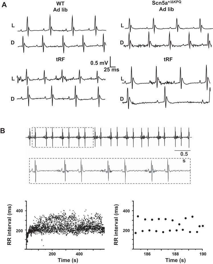
Fig. 4.
Light phase-restricted feeding unmasks ECG differences between WT and Scn5a+/ΔKPQ mice. A: representative ECG traces recorded in WT (left) and Scn5a+/ΔKPQ (right) mice during both light (L) and dark (D) phase in ad libitum (top 2 traces)- or tRF-feeding (bottom 2 traces) conditions. B: small portion of an atrial bigeminy arrhythmia recorded from an Scn5a+/ΔKPQ mouse. Inset: boxed ECG region at a higher resolution. Graphs show beat-to-beat changes in the RR interval measured during on entire episode of a bigeminal arrhythmia (left) or an amplified portion (right) to highlight the alteration in the RR intervals.

