
Fig. 11.
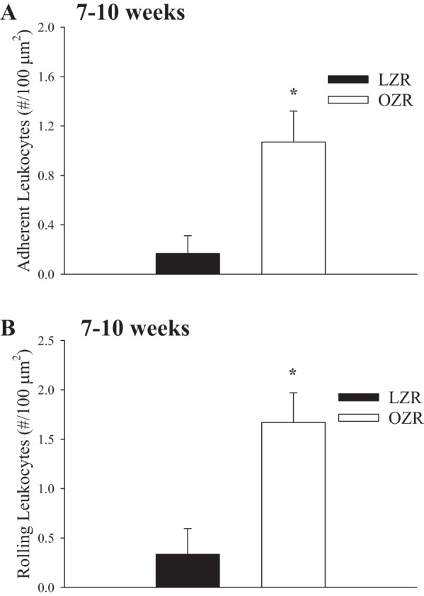
Data describing the adhesion (A) and rolling (B) of leukocytes in the postcapillaries of venules of in situ extensor digitorum longus muscle of LZR and OZR at 7–10 wk of age. Rhodamine 6G-labeled vascular cells were visualized by epi-illumination. The number of rolling and adherent leukocytes in venules was expressed as number of cells/are of endothelial surface with the denominator calculated from the length and diameter of the venular segment. *P < 0.05 vs. LZR.