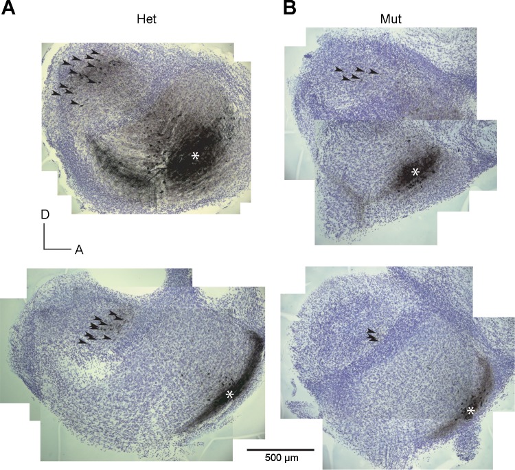
Fig. 4.
Topographic organization of intrinsic cochlear nuclear connections. Photomicrographs of sections from 4 slices show location of injections of biocytin into the anteroventral cochlear nucleus (aVCN, *). The injections labeled a discrete bundle of auditory nerve fibers and a group of tuberculoventral neurons in the deep layer of the DCN; as these labeled neurons are difficult to resolve at low power, they are marked with arrowheads. A: in heterozygotes, injections into the ventral aVCN label tuberculoventral neurons ventrally (bottom) and more dorsal injections label neurons more dorsally (top). B: labeled tuberculoventral neurons in Otoferlin mutant mice follow the same pattern. The more ventral injections label tuberculoventral cells more ventrally than the most dorsal injections. Top left panel is a composite image of photomicrographs of 2 sections; all other panels are composite images of individual sections.

