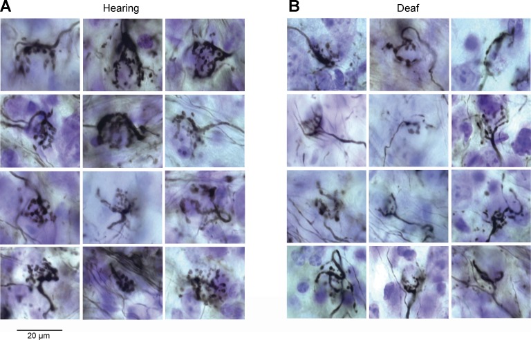
Fig. 5.
Auditory nerve fibers and their end bulb terminals. Photomicrographs of end bulbs labeled with extracellular injections of 1% biocytin into the nerve root, aVCN, or posteroventral cochlear nucleus (pVCN). A: end bulbs from hearing mice comprise highly branched clusters of boutons that engulf the cell body of target bushy cells. B: end bulbs from deaf Otoferlin mutant mice are smaller. In some, the axons that lead to the end bulbs are small and thin.

