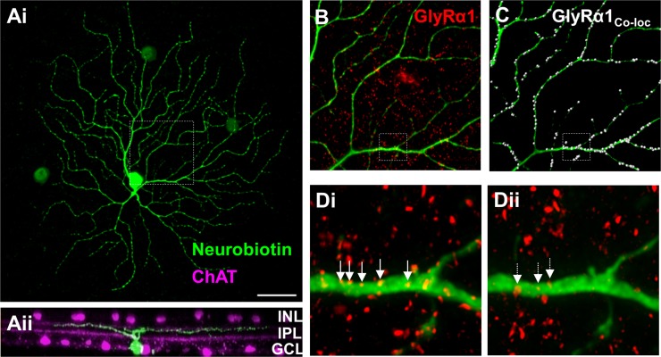
Fig. 1.
The GlyRα1 subunit is expressed on PV5WT retinal ganglion cell (RGC) dendrites. Ai: representative confocal image of a neurobiotin-labeled PV5 RGC (green) in a retinal whole mount shows its characteristic A2 type morphology. ChAT, choline acetyltransferase. Aii: rotated view of the same RGC shows its dendritic lamination pattern in the Off sublaminae of the inner plexiform layer (IPL) relative to the bands formed by the processes of the cholinergic amacrine cells (ACs; magenta). B: representative portion of the dendritic arbor of the same PV5WT RGC (see box; 70 × 70-μm area) and the punctate pattern of GlyRα1 subunit expression (red). C: colocalized GlyRα1 puncta (white dots) on the dendrite of the same RGC. Di: magnified and deconvolved image from B (see box). Arrows indicate a subset of representative colocalized GlyRα1 puncta on the PV5 dendrite. Dii: illustration of random association of GlyRα1 puncta that results from superposition of the channel containing the dendrite with a duplicated and 180°-rotated channel with the GlyRα1 puncta. Arrows indicate what are considered randomly colocalized GlyRα1 puncta. The number of randomly colocalized puncta was used to correct GlyRα1 puncta coincidence rate. INL, inner nuclear layer; GCL, ganglion cell layer. Scale bar (shown in Ai): 40 μm (A), 16 μm (B and C), 2.5 μm (D).

