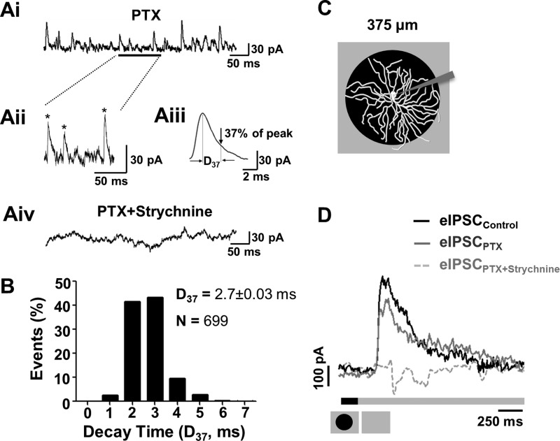
Fig. 2.
GlyRα1-meditated spontaneous inhibitory postsynaptic currents (sIPSCs) and glycinergic stimulus-evoked IPSCs (eIPSCs) are present in PV5WT RGCs. Ai: glycinergic sIPSCs were recorded from PV5WT RGCs held at 0 mV in the presence of picrotoxin (PTX; 20 μM). Aii: same trace as Ai with an expanded timescale. Asterisks indicate sIPSCs that met our criterion (see text) and were used in our analyses. Aiii: average waveform of PV5 RGC sIPSCs that met our criterion (170 events) and an illustration of their average decay (time at which sIPSC declined to 37% of peak amplitude, D37). Aiv: all sIPSCs were eliminated in presence of PTX (20 μM) and strychnine (10 μM). B: frequency of decay times (D37) in sIPSCs of PV5WT RGCs (n = 12; N = 699 events). C: schematic representation of the visual stimulus, a dark spot (6,000 R*·rod−1·s−1) centered on the PV5WT RGC soma and presented on a background (24,000 R*·rod−1·s−1). A spot outer diameter of 375 μm matches the PV5 RGC dendritic arbor. D: averaged PV5WT RGC eIPSC evoked by the offset of the dark spot in control solution. PTX (20 μM) produces a small reduction in the eIPSC, indicating a small GABAergic input. The eIPSC is completely eliminated when strychnine also is present (PTX 20 μM + strychnine 10 μM), indicating that the majority of synaptic inhibitory input is glycinergic.

