Figure 1.
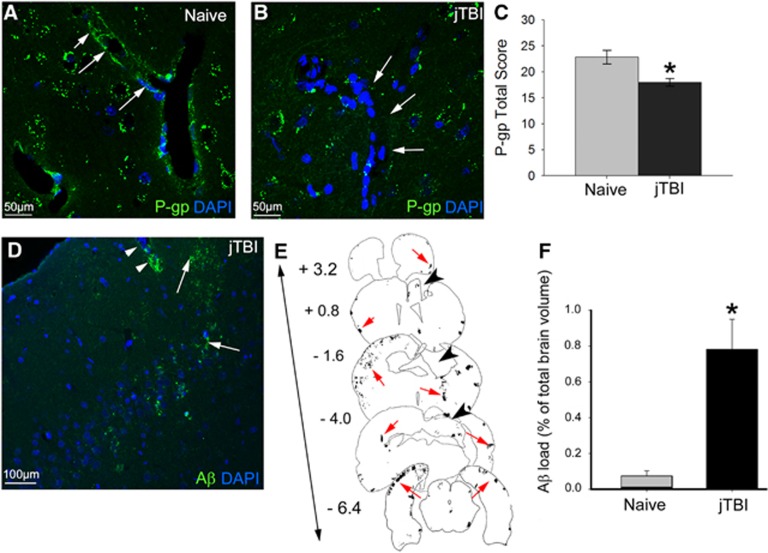
Juvenile traumatic brain injury (jTBI) induces amyloid-beta (Aβ) accumulation and decrease of P-glycoprotein (P-gp) levels in the brain 6 months after injury. (A and B) P-glycoprotein staining was observed in endothelial cells of cortical vessels in both naive and jTBI animals (arrows). (C) Quantification of P-gp in the cortex shows a significant decrease in jTBI animals compared with naive (*P<0.05). (D) Rodent-Aβ was detected in diffuse extracellular deposits (arrowheads) and around blood vessels (arrows) 6 months after injury. (E) Representative coronal sections with outlines of rodent-Aβ staining (red arrows) are shown at bregma +3.2, +0.8, −1.6, −4.0, and −6.4 mm in a jTBI animal 6 months after injury. The lesion cavity is apparent at bregma +0.8, −1.6, and −4.0 mm (black arrowheads). (F) Aβ load is significantly higher in jTBI animals compared with naive (*P<0.05). Scale bars, 50 μm (A and B); 100 μm (D).

