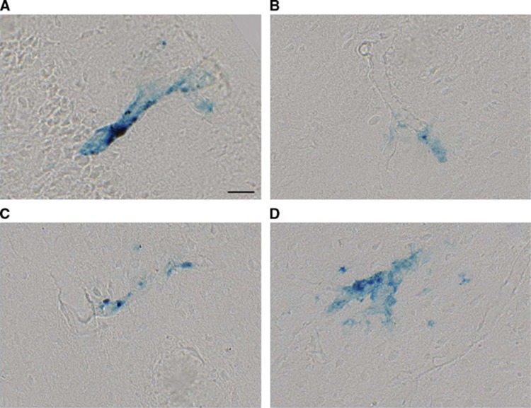
Figure 3.
Cortical microhemorrhages were detected by Perls Prussian blue staining. Representative vascular leaks identified in insular (A and B), secondary motor (C) and primary motor (D) cortices. Scale bar=25 μm. Examples shown are from an APP+ >14Mo mouse, and representative of observations in APP+ mice in general.

