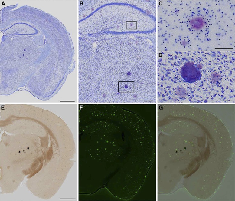
Figure 5.
(A) Coronal sections from APP+ mice were stained with cresyl violet to evaluate histopathology areas where thalamic computed tomography (CT) hyperintensities were detected. Thalamic deposits were easily identified by their deep purple color with this stain. (B–D) At higher magnification, amyloid plaques display a characteristic starburst shape in Nissl-stained tissue (C) while calcospherites are distinguished by their globular morphology (D). (E–G) Serial tissue sections were stained with von Kossa's method (E) and thioflavine-S (F) to evaluate coincidence of calcospherites with amyloid. Mineralizations appear black in von Kossa-stained sections and do not overlap with thioflavine-S-stained plaques in green (G). Scale bars: (A and E) 1 mm; (B) 500 μm; (C and D) 50 μm. Examples shown are from an APP+ >14Mo mouse, and representative of observations in APP+ mice in general.

