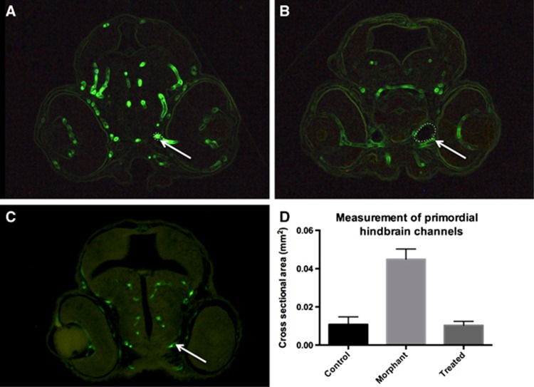
Figure 3.
Histologic assessment of cerebral vasculature shows dilated cranial blood vessels in an arteriovenous malformation (AVM) model. 4-dpf zebrafish were embedded in JB-4 resin, serially sectioned, and visualized at the level of the mid-eye in the coronal plane. Measurements of the primordial hindbrain channels were significantly smaller in the (A) uninjected organisms compared with the (B) morphant-control group (P<0.0001). (C, D) Morphant-treatment group measurements (0.0103±0.00101 mm2) were not significantly different from uninjected organisms (P=0.8574), consistent with a rescue phenomenon of the vascular phenotype.

