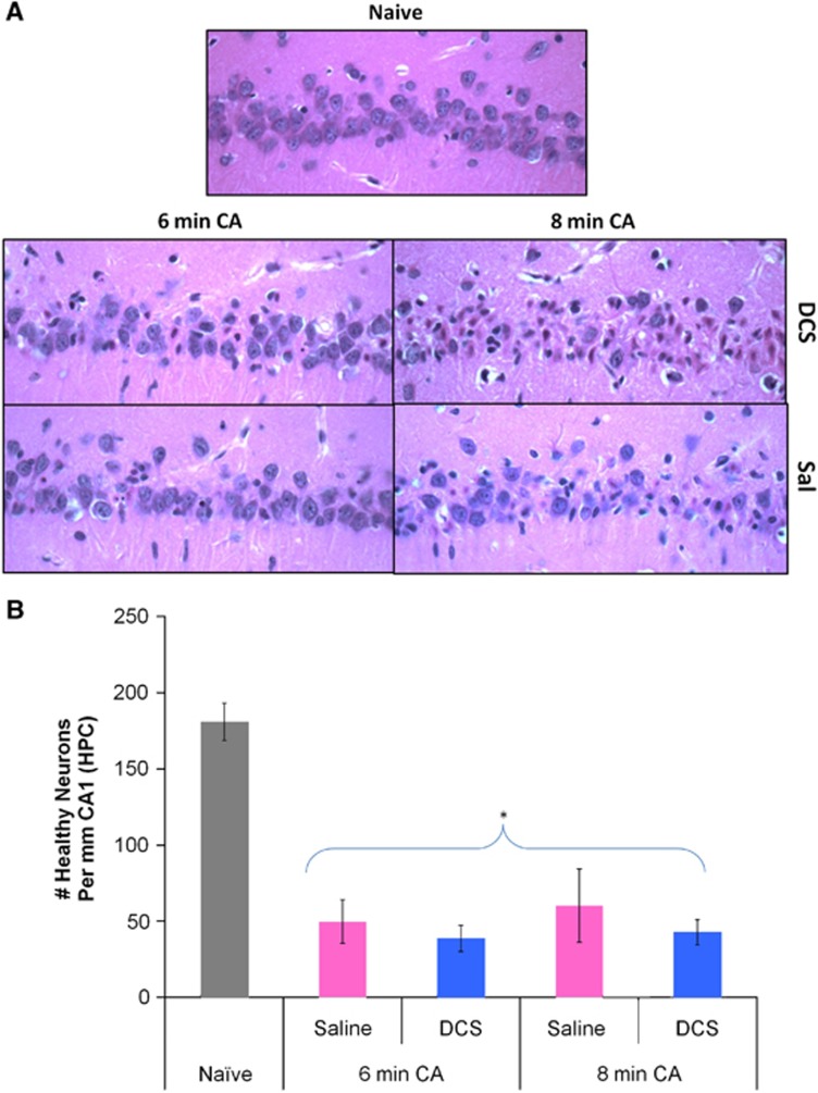
Figure 6.
(A) Representative images (at 40X magnification) showing pyknotic (ischemic) and healthy neurons in the CA1 hippocampal region of drug (D-cycloserine; DCS) or Vehicle (Sal) treated rats 8 days after 6 minutes or 8 minutes of asphyxial cardiac arrest (CA). (B) Number of healthy neurons per mm of hippocampal (HPC) CA1 in naïve rats and rats subjected to 6 minutes or 8 minutes of CA and treated with saline or DCS. No significant differences in CA1 neuronal cell counts were noted between experimental groups; however, neuronal counts in all CA groups differed from naïve rats. *P<0.05 CA, n=27 versus naïve, n=5, t-test; mean±s.e.m.

