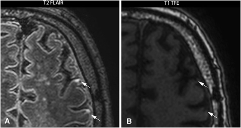
Figure 1.
7.0 Tesla T2-weighted fluid-attenuated inversion recovery (FLAIR) image (A) and a T1-weighted turbo field echo (TFE) image (B) of a 74-year-old male with a symptomatic high-grade stenosis of the left carotid artery scheduled for carotid endarterectomy. Two cortical microinfarcts (arrows) ipsilateral to the symptomatic carotid artery are visualized in transversal plane.

