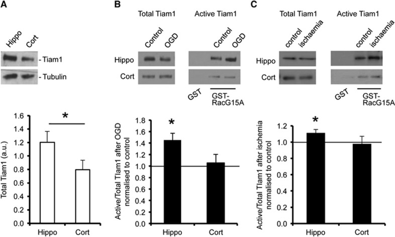
Figure 3.
Tiam1 is activated in response to oxygen/glucose deprivation (OGD) in hippocampal, but not in neurons. (A) Endogenous expression of Tiam1 (~177 kDa) is higher in hippocampal neurons compared with cortical neurons. Representative blots show total endogenous Tiam1 in cell lysates prepared from hippocampal and cortical cultures. Tubulin (~55 kDa) was the loading control. Graph shows Tiam1 level normalized to tubulin presented as mean±s.e.m. *P<0.05 (T-test). Hippocampal neurons, n=6 independent cultures; cortical neurons, n=6 independent cultures. (B) Cell lysates from control conditions or after 20 minutes of OGD were incubated with GST-RacG15A immobilized on glutathione-agarose beads to isolate activated Tiam1. Cell lysates from control conditions were also incubated with GST as a negative control. Representative blots show the levels of active (RacG15A-bound) and total Tiam1 in hippocampal and cortical neurons under control and OGD conditions. Graph shows pooled data presented as mean±s.e.m. The proportion of active Tiam1 increased in hippocampal neurons but was unchanged in cortical neurons in response to OGD. *P<0.05 (T-test). Hippocampal neurons, n=8 independent cultures; cortical neurons, n=7 independent cultures. (C) Lysates from cortical and hippocampal synaptoneurosomes prepared from rats subjected to transient forebrain ischemia or controls were incubated with GST-RacG15A immobilized on glutathione-agarose beads to isolate activated Tiam1. Control samples were also incubated with GST as a negative control. Representative blots show the levels of active (RacG15A-bound) and total Tiam1 in hippocampal and cortical synaptoneurosomes under control and ischemic conditions. Graph shows pooled data presented as mean±s.e.m. The proportion of active Tiam1 increased in hippocampal synaptoneurosomes but was unchanged in cortical neurons in response to ischemia. *P<0.05 (T-test). Control, n=7 animals; ischemia, n=8 animals. GST, Glutathione-S-transferase.

