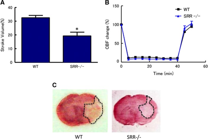
Figure 1.
Reduced infarct volumes in serine racemase (SRR) −/− mice. (A) Infarct volume was determined 24 hours after middle cerebral artery occlusion (MCAO) (n=7 to 8/group); *P<0.05 vs. wild type (WT) (Student's t-test). (B) Core blood flow during and after MCAO (n=7 to 8/group); not significant between WT and SRR−/− (analysis of variance, ANOVA). (C) Representative triphenyltetrazolium chloride-stained brain sections derived from animals analyzed in (A). CBF, cerebral blood flow.

