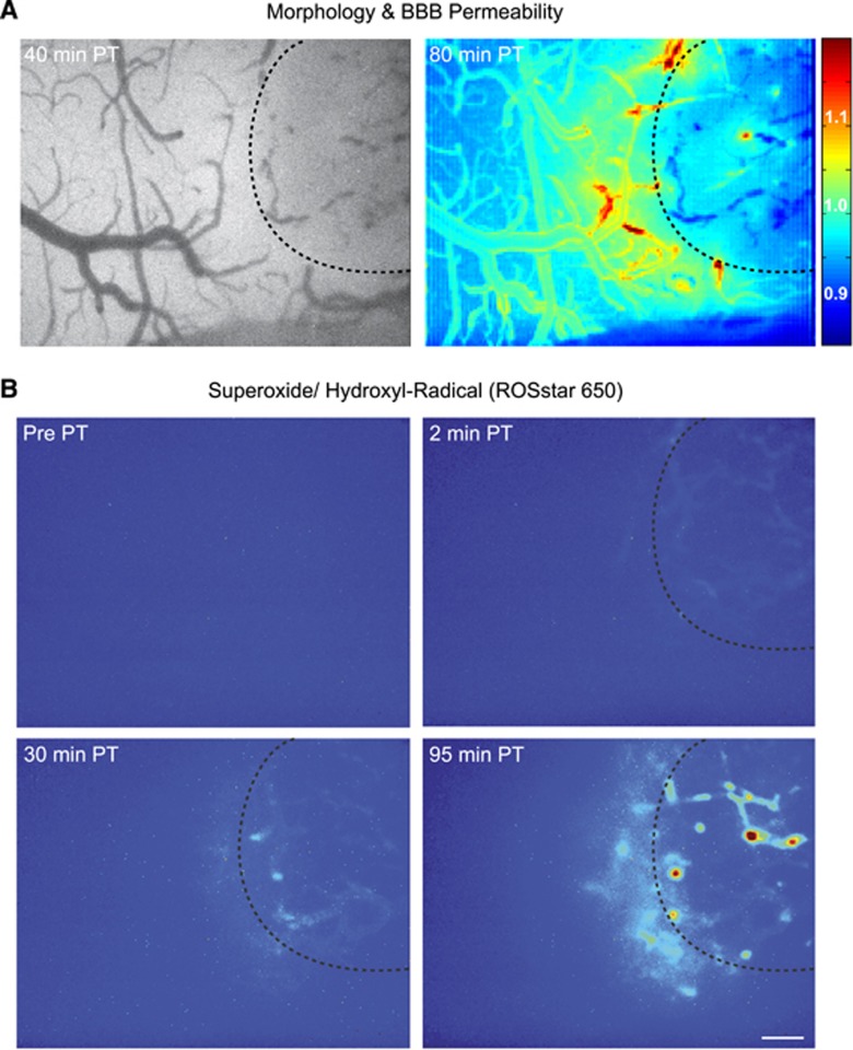
Figure 4.
Intravital microscopy reveals superoxide and hydroxyl radical formation after photothrombosis (PT). (A) Left: bright field image 40 minutes after PT induction. Right: blood–brain barrier (BBB) permeability index 80 minutes after PT. (B) Areas with enhanced superoxide and hydroxyl radical formation. Note the detection of free radicals in the ischemic core as well as in the peri-ischemic cortex. (A and B) Dotted lines indicate previously laser-illuminated ischemic core. Scale bar=200 μm.

