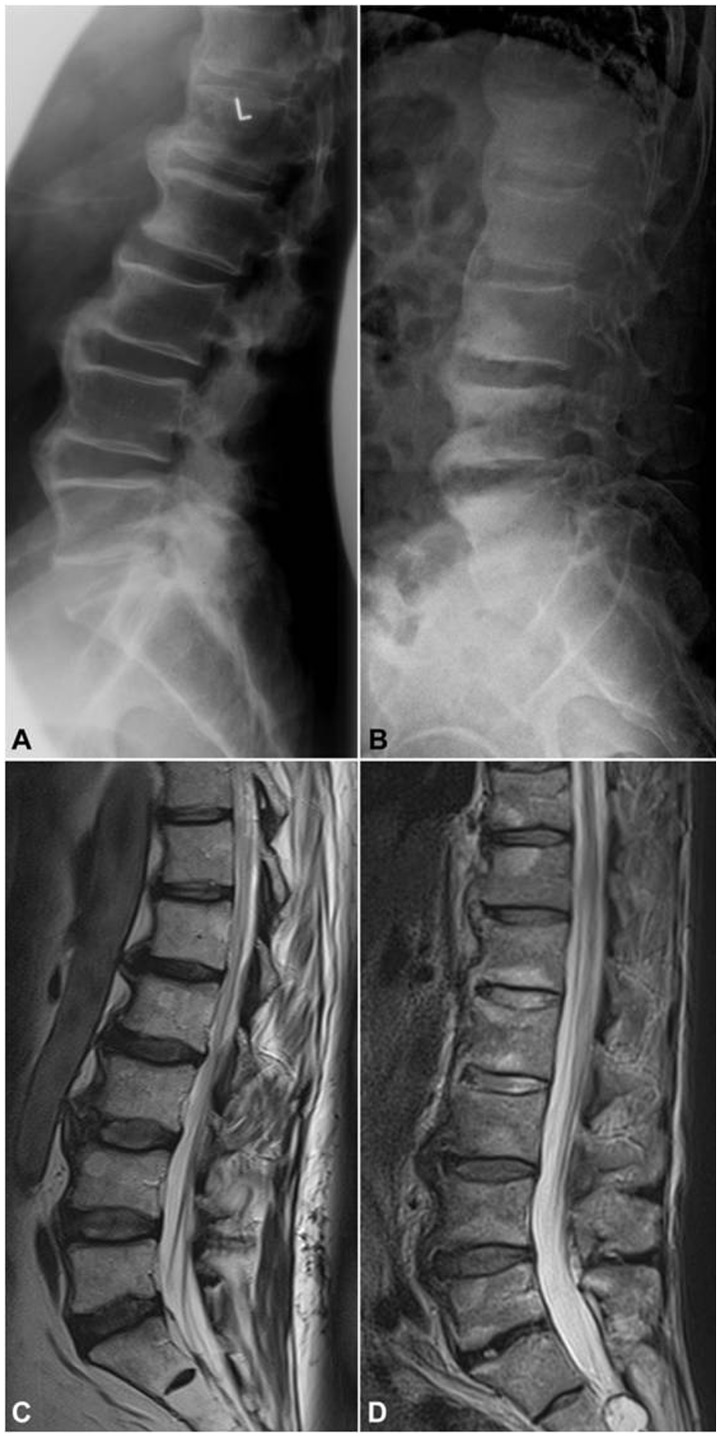
Fig. 3.
Spinal similarities in OA and PsA as seen on imaging
A 69-year-old male with chronic low back pain and gradual stiffening of the spine showing excessive osteophytosis at the lumbar spine as depicted by (A) lateral X-ray and (C) T2-weighted sagittal MRI, compatible with diffuse idiopathic skeletal hyperostosis (DISH), which is an OA variant. In contrast, the 65-year-old male depicted by (B) X-rays and (D) T2-weighted MRI suffers from long-standing psoriatic spondylitis and presents with so called parasyndesmophytes along with inflammatory discal lesions at the lumbar spine. Of note, disc height is well preserved in images of both patients. (B) and (C) courtesy of Dr S. Hermann, Rheumatology, Charité Medical School, Berlin, Germany.

