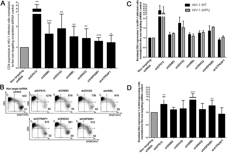
Figure 3.

Effect of the knock-down of the selected genes on CD4 expression in HIV-1-infected SupT1 cells. A. The bar graph shows the residual CD4 surface expression levels in HIV-1-infected (eGFP+) SupT1 cells (calculated as described in Methods) normalized to the non-targeting shRNA scrambled control. Error bars represent standard deviation between 7 to 17 independent experiments. The values are representative of the shRNA clone with most activity among the ones tested. P values: ***p ≤ 0.0004; **p ≤ 0.0099; *p ≤ 0.038. B. Flow cytometry dot plots showing the CD4 surface levels in HIV-1-infected cells in a representative experiment. eGFP is the reporter for HIV-1 infection, while CD4 levels are measured by surface staining with an anti-CD4 monoclonal antibody conjugated to APC. The numbers on the FACS plots indicate the mean fluorescence intensity of CD4 gated on the upper right and lower right quadrants. C. The bar graph shows the residual CD4 surface expression levels in shRNA expressing SupT1 cells normalized to the non-targeting shRNA scrambled controls (calculated as described in Methods), infected with the wild type (black bars, indicated as HIV-1 WT) and the dVpu HIV-1 NL4-3 IRES-EGFP (grey bars, indicated as HIV-1 dVpu). Error bars represent standard deviation between 3 independent experiments. D. The bar graph shows the residual CD4 surface expression levels (calculated as described in Methods) in SupT1 cells transduced by a retroviral vector to express Nef, normalized to the non-targeting shRNA scrambled control. Error bars represent standard deviation between 7 to 16 independent experiments. P values: ***p ≤ 0.0006 ; **p ≤ 0.0011.
