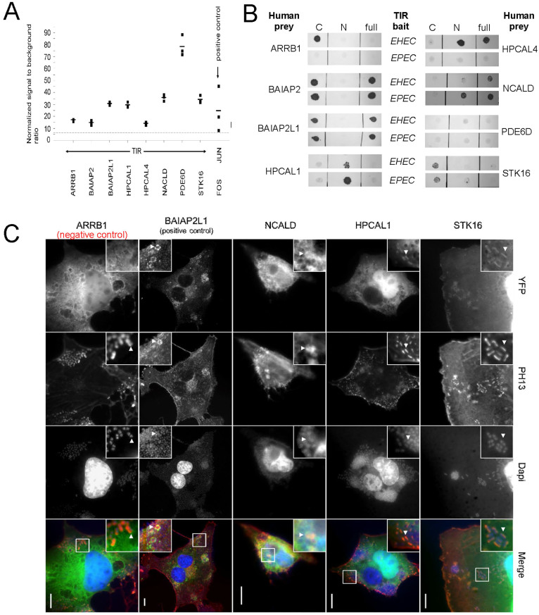
Figure 5. Validation and conservation of TIR-host interactions.
(a) TIREHEC interactions that were detected in our Y2H screens were confirmed by LUMIER assays, using full-length TIR as protein A fusion and the human test partners co-purified as luciferase-tagged fusions. Black squares represent individual measurements of the co-purified luciferase by luminescence, while their averages are shown as black horizontal bars. We considered values above the dashed threshold line (signal to background ratio >6) as a positive binding signal. As a positive control we used JUN/FOS. (b) Comparison of homologous interactions of TIR in EHEC and EPEC by pairwise Y2H tests. In particular, we probed full-length constructs (full) as well as the N- and C-terminal cytosolic domains (not shown) as baits against human preys. (c) We tested the co-localization of TIR interactors with EPEC infection sites on COS-7 cells and used BAIAP2L1 that interacts with TIR as positive control. Co-localization sites are indicated by white arrows. Specifically, we fused (YFP) human binders C-terminally to YFP and used (PH13) F-actin staining to visualize pedestals and (Dapi) DNA staining.

