
Figure 1.
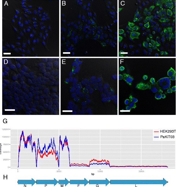
HeV infection and transcription in bat and human cells. Confocal microscopy was used to visualize HeV-infected cells. Nuclei are stained with 4′,6-diamidino-2-phenylindole dihydrochloride, and HeV-N protein was immunodetected with an anti-N polyclonal antibody. PaKiT03 cells at (A) 0 hpi, (B) 8 hpi and (C) 24 hpi are shown. HEK293T cells infected with HeV for (D) 0 hpi, (E) 8 hpi and (F) 24 hpi are shown. Scale bar is 30 μm. (G) Transcription profile of HeV in PaKiT03 and HEK293T at 24 hpi. (H) Genome structure of HeV. bp, base pairs; F, fusion protein; G, glycoprotein (attachment protein); L, large protein (polymerase); M, matrix protein; N, nucleocapsid; P, phosphoroprotein (includes proteins V, W and C).