
Figure 7.
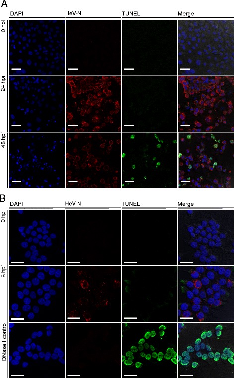
TUNEL staining of HeV-infected (A) PaKiT03 and (B) HEK293T cells. Cells were infected with HeV for either 8, 24 or 48 h using an MOI of 5. TUNEL staining was achieved using the Click-iT® TUNEL Alexa Fluor® 488 kit (green, fluorescence) and HeV-N was immunodetected as described above (red, fluorescence). DNase I treatment was used as a positive control on HEK293T cells that demonstrated clear nuclear staining. Owing to the decrease in cell viability at 24 hpi and beyond in the HEK293T cells, reliable TUNEL staining could not be performed after 8 hpi. Scale bar is 30 μm in all panels.