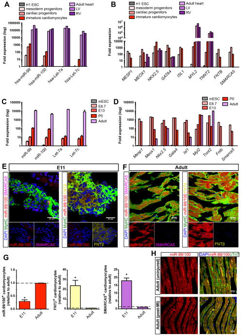
Figure 4. The miR-99/100-dependent heart regeneration pathway is developmentally conserved in mammals but fails to activate upon injury.
(A–B) qRT-PCR analysis showing the expression of miR-99/100 and Let-7a/c (A), their protein targets and key cardiac transcription factors (B) during human cardiac differentiation (n=5). Human embryonic stem cell line H1 (H1 ESC); left ventricle (LV and right ventricle (RV). (C–D) qRT-PCR analysis showing the expression of miR-99/100 and Let-7a/c (C), their protein targets and key cardiac transcription factors (D) during mouse development and adult stages (n=5). Mouse embryonic stem cells (mESC). (E–G) FISH/immunofluorescence (E, F) and quantitative analysis (G) demonstrating low levels of miR-99/100 and high levels of FNTβ and SMARCA5 in E11 murine embryonic hearts as opposed to adult hearts, which showed high levels of miR-99/100, and almost undetectable expression of FNTβ and SMARCA5. (H) FISH/immunofluorescence in adult mouse heart before (upper panels) and after myocardial infarction (lower panels) highlighted a failure to downregulate miR-99/100 upon injury in the murine heart. Data are represented as mean ± s.e.m. *p<0.05. (E, F): n = 6 animals; (H): n= 5 animals. In all cases, three different sections per animal were used for quantitative analyses (G). See also Figure S4.

