
Fig. 5.
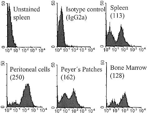
Differential expression of CD38 on B lymphocytes from different lymphoid compartments. Total lymphocytes from spleen, peritoneal cavity, Peyer's patches and bone marrow were stained with FITC-anti-mouse CD38 (NIM-R5). Numbers in parentheses show the mean fluorescence intensity of the CD38+ cells.