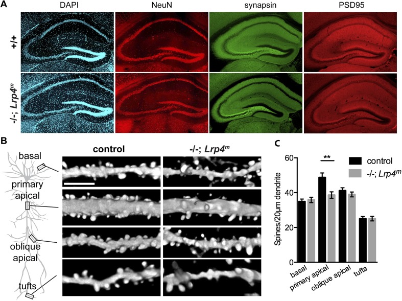
Figure 8. A loss of Lrp4 decreases spine density in primary apical dendrites of CA1 neurons.
(A) The organization of the hippocampus appears normal as assessed by staining sections of the adult hippocampus for DNA (DAPI), neuronal nuclei (NeuN), presynaptic terminals (synapsin) or excitatory postsynaptic membranes (PSD95). (B) Representative images of dendrites of Thy1-YFP labeled CA1 Lrp4 mutant pyramidal neurons near (basal and apical) and far (oblique apical and tufts) from the cell body. Scale bar: 5 μm. (C) The spine density is reduced selectively at primary apical dendrites (basal: wild-type, n = 45; Lrp4−/−; Lrp4m, n = 45; primary apical: wild-type, n = 43; Lrp4−/−; Lrp4m, n = 46; oblique apical: wild-type, n = 42; Lrp4−/−; Lrp4m, n = 46; tufts: wild-type, n = 42; Lrp4−/−; Lrp4m, n = 45).

