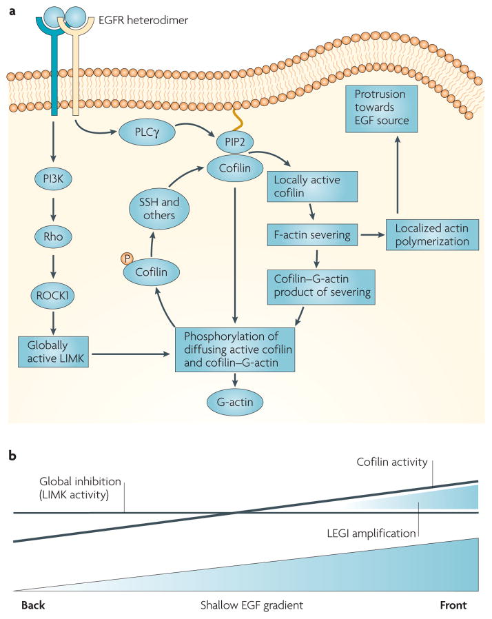
Figure 2. The spatial and temporal localization of cofilin activity in response to EGF stimulation.
a | In vivo studies in tumour cells indicate that EGF-stimulated phospholipase Cγ (PLCγ) hydrolyses phosphatidylinositol-4,5-bisphosphate (PIP2), causing the release of active cofilin locally from its complex with PIP2 in the plasma membrane. This activates cofilin asymmetrically inside the tumour cell to generate free barbed ends adjacent to the cell membrane facing the source of epidermal growth factor (EGF). Transient cofilin activity occurs as the result of the near simultaneous activation of the molecules within the cofilin pathway that both activate cofilin (PLCγ, slingshot (SSH) and chronophin) and inhibit cofilin (LIMK) by the EGF receptor. As a result, cofilin severs filaments locally to start polymerization and cell protrusion, and global LIMK activity inactivates cofilin that diffuses from the initial site of activation, thereby spatially sharpening cofilin activity. The signalling pathways from the receptor through G-proteins and LIMK are the same as in FIG. 1 but redrawn here to show how LIMK captures cofilin that diffuses from the site of activation. This results in the sharpened localization of cofilin-dependent free barbed end production and the initiation of directional cell motility and chemotaxis. b | The cofilin activity cycle fits a local excitation global inhibition (LEGI) model of chemotaxis. Cofilin is activated asymmetrically inside the cell in response to the gradient in EGF detected by cell surface receptors from the front to the back of the cell. The asymmetry of cofilin activation inside the cell may follow the slope of the gradient of EGF. However, this shallow gradient in cofilin activation is locally sharpened by the global stimulation of LIMK activity, which is postulated to inactivate cofilin throughout the cell, resulting in a remnant of cofilin activity only on the side of the cell facing the EGF source (front). This is shown by the two lines indicating the cofilin activation gradient (sloping line) and global LIMK activity (horizontal line) resulting from the same stimulation with a shallow EGF gradient, thereby compressing the effective cofilin activity between them to sharpen cofilin activity to the side of the cell facing the EGF source.

