. 2014 Nov 11;3:e04132. doi: 10.7554/eLife.04132

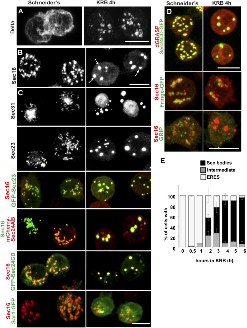
Figure 1. Amino-acid starvation induces the formation of a novel stress assembly in Drosophila S2 cells.

(A) Immunofluorescence (IF) visualization of Delta-myc (using an anti-Delta antibody) in S2 cells in Schneider's (normal growth conditions) or incubated with Krebs Ringer Bicarbonate buffer (KRB) for 4 hr (amino-acid starvation). Note that in Schneider's Delta reaches the plasma membrane whereas it is retained intracellularly in starved cells. (B) IF visualization of endogenous Sec16 in Drosophila S2 cells grown in Schneider's and incubated in KRB for 4 hr. Note the formation of Sec bodies (arrows). (C) IF visualization of Sec31, Sec23 and co-visualization of GFP-Sec23, mCherry-Sec24AB, Sec24CD-GFP, Sar1-GFP with Sec16 in S2 cells in Schneider's and KRB for 4 hr. (D) IF co-visualization of dGRASP/Sec24CD-GFP, GRIP/Sec16, and Fringe-GFP/Sec16 in S2 cells grown in Schneider's and incubated in KRB for 4 hr. (E) Kinetics of Sec body formation in S2 cells incubated in KRB over indicated time (up to 6 hr) expressed as the percentage of cells exhibiting ERES, intermediates (see ‘Materials and methods’), and Sec bodies. Scale bars: 10 μm (AD).

DOI: http://dx.doi.org/10.7554/eLife.04132.003

Figure 1.

Figure 1—figure supplement 1. Sec body formation and autophagy.

Figure 1—figure supplement 1.

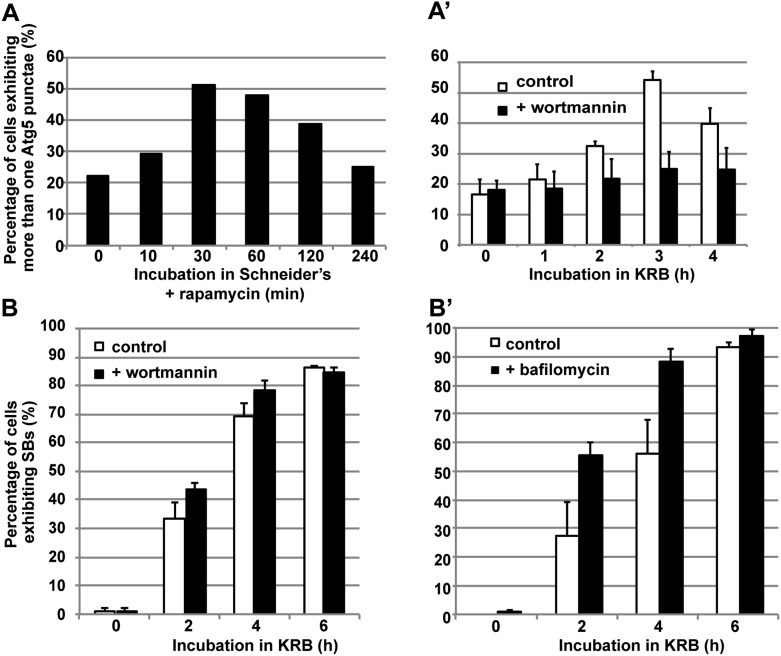
(AA′) Quantification of Atg5 punctae formation in S2 cells incubated with rapamycin (A) and incubated with KRB with or without wortmannin (A′) for indicated time points. Note that as expected, autophagy (marked by Atg5) is stimulated by rapamycin and starvation (KRB) and inhibited by wortmannin. (BB′) Quantification of Sec body formation (marked with Sec16) in cells incubated in KRB with and without wortmannin (B) and with and without bafilomycin (B′).

Figure 1—figure supplement 2. Sec body formation and single amino-acids.

Figure 1—figure supplement 2.

Quantification of the prevention of Sec body formation by specific amino-acids upon incubation in KRB for 4 hr. This is expressed as the percentage of cells exhibiting the normal growth Sec16 localization at ERES. All amino-acids are added at 15 mM except for 3 (gray bars) that were also tested at their concentration in Schneider's medium. Note that at 15 mM, histidine, aspartate, and asparagine significantly decreases Sec body formation.

Figure 1—figure supplement 3. Sec body formation in vivo.

Figure 1—figure supplement 3.

Projection of four equatorial confocal planes of Sec16 and Spectrin (D) in the follicular epithelium covering an egg-chamber from an ovary dissected from a virgin fly fattened for 3 days and incubated ex-vivo in KRB for 4 hr, and from an ovary dissected from a 36-hr starved virgin female. Note that in both cases, Sec16 is found in large punctae reminiscent of Sec bodies. Scale bars: 10 μm.

Figure 1—figure supplement 4. Sec body formation and mammalian cells.

Figure 1—figure supplement 4.

IF visualization of Sec16 in immortalized MEFs incubated in growth medium (DMEM) or KRB plus bafilomycin for 7 hr. Note that Sec16 is remodeled into larger structures. Scale bar: 10 μm.

Figure 1—figure supplement 5. Human Sec16A is incorporated to Sec bodies in starved Drosophila S2 cells.

Figure 1—figure supplement 5.

IF visualization of human Sec16A-V5 transfected in Drosophila S2 cells. Note that it partially localizes to ERES in fed cells but is efficiently incorporated in Sec bodies upon starvation. Scale bar: 10 μm.