. 2014 Nov 11;3:e04132. doi: 10.7554/eLife.04132

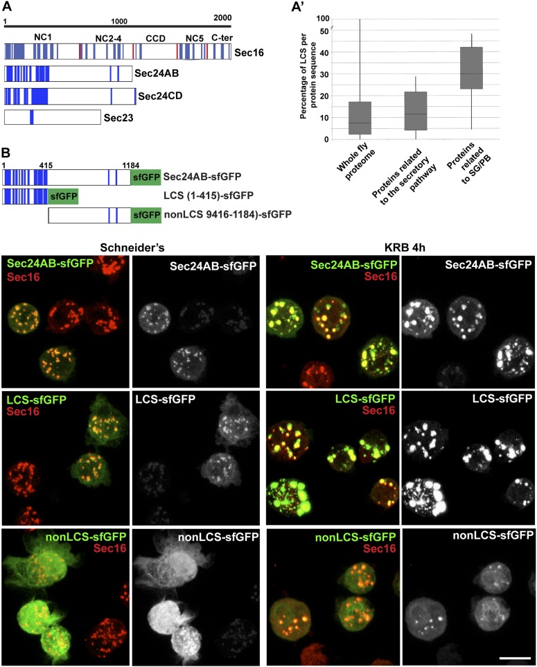
Figure 6. Sec body proteins contain low complexity sequences that are necessary for Sec body recruitment.

(AA′) Schematic representation of the Low Complexity Sequences (blue bars) in Sec16, Sec24AB, Sec24CD, and Sec23 (A). The red bars mark the boundaries of the Sec16 domains. Genome wide analysis of Low Complexity Sequence (LCS) in the Drosophila proteome, in proteins related to the secretory pathway and proteins related to Stress Granules/P-bodies (A′). B: IF localization of sfGFP-tagged full-length Sec24B, Sec24AB LCS, and Sec24AB nonLCS in S2 cells in Schneider's and KRB for 4 hr, together with endogenous Sec16 (red). Scale bars: 10 μm.

DOI: http://dx.doi.org/10.7554/eLife.04132.015

Figure 6—Source data 1. Table showing the LCS content of proteins related to Stress Granules and P-bodies as well as the early secretory pathway (related to Figure 6A,A′).
DOI: 10.7554/eLife.04132.016

Figure 6.

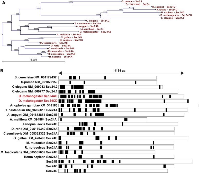
Figure 6—figure supplement 1. LCS content of Sec24 in different species.

Figure 6—figure supplement 1.

(A) A restricted phylogenic tree of the Sec24 sequences analyzed in (B) Note that S. pombe and S. cerevisiae Sec24 are similar to each other and to H. sapiens Sec24C and D, and D. melanogaster Sec24CD. However, they are distant to D. melanogaster Sec24AB, H. sapiens Sec24A and B. Furthermore, D. melanogaster Sec24AB is distant to D. melanogaster Sec24CD. (B) LCS analysis and schematics in Sec24 sequences of different organisms. Note that most sequences contain a significant percentage of LCS in the N-terminal third of the protein with the exception of S. pombe, A. melifera and G. gallus (related to Figure 6A).
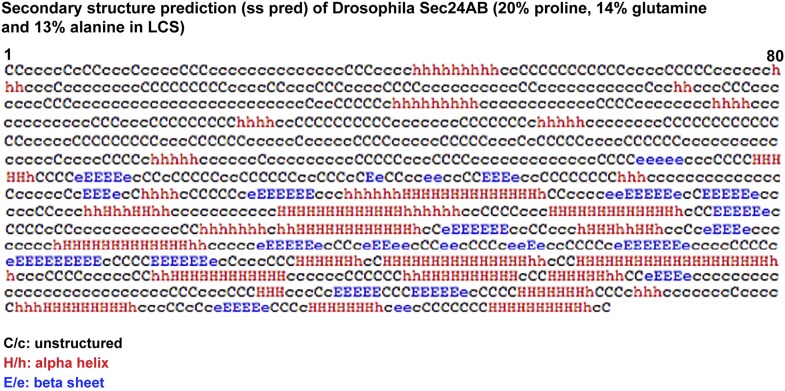
Figure 6—figure supplement 2. Secondary structure prediction of Drosophila Sec24AB using HH pred.

Figure 6—figure supplement 2.

C/c denotes the unstructured, H/h the alpha helices, and E/e, the beta sheets. Note their absence in the 405 amino-acids of the N-terminus corresponding to LCS.
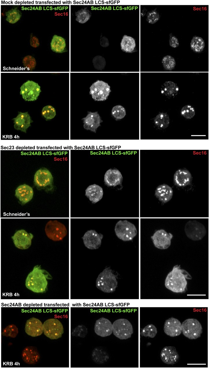
Figure 6—figure supplement 3. Sec24AB LCS is not sufficient to drive Sec body formation.

Figure 6—figure supplement 3.

S2 cells were depleted of endogenous Sec24AB. When starved (KRB), this resulted in the formation of small structures (as in Figure 5). The transfection of Sec24AB LCS-sfGFP in these depleted cells did not rescue the formation of Sec bodies. Scale bars: 10 μm.