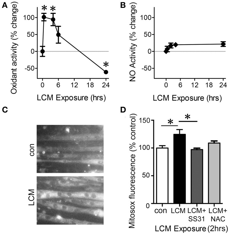
Figure 4.
LCM treatment alters oxidant production. (A) DCFH fluorescence, a measurement of cytosolic oxidant levels, was increased by LCM treatment for 30 min and 4 h, but reduced by 24 h of treatment, represented as percent change (n = 10, *P < 0.05, ANOVA, Bonferroni post-hoc). (B) Unaltered DAF fluorescence in myotubes treated with LCM for 30 min, 2, and 24 h, represented as percent change (n = 20, non-significant, ANOVA, Bonferroni post-hoc). (C) Fluorescence images of control myotubes (top panel) and LCM-treated myotubes (bottom panel) treated with MitoSox. (D) 2 h of LCM treatment increased MitoSox fluorescence compared to control; 2 h of LCM treatment combined with SS31 decreased Mitosox fluorescence compared to LCM-treated myotubes; and 2 h of LCM treatment combined with NAC did not alter MitoSox fluorescence significantly (n = 4, *P < 0.05, Bonferroni post-hoc).

