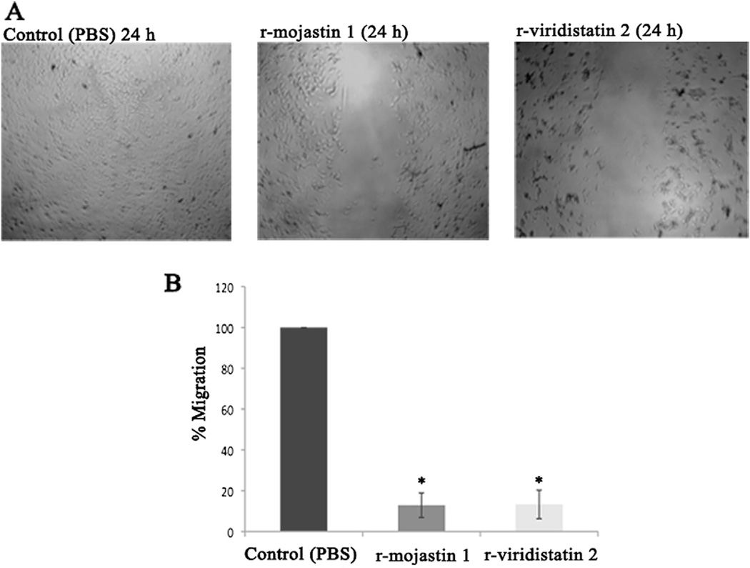
Fig. 3.
Migration of HUVECs in presence of r-viridistatin 2 and r-mojastin 1 (A & B). HUVECs (9 × 10⁴ cells/mL) were seeded in 48-well plates in the presence of r-viridistatin 2 and r-mojastin 1 at 6.3 µM for 24 h. The negative control consisted of cells treated with PBS buffer, pH 7.4. The results are expressed as the percentage of cell migration relative to the negative control. The results are expressed as mean ± SD (n = 3). *p < 0.05 when compared to control (PBS).

