
Figure 2.
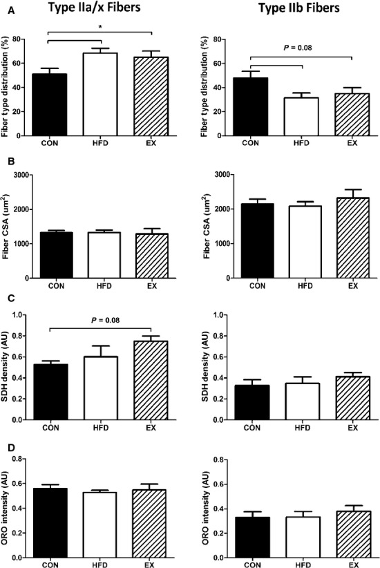
Tibialis anterior morphology. (A) Fiber‐type distribution based on myosin heavy chain isoform staining. *HFD versus CON. (B) Fiber cross‐sectional area. (C) Succinate dehydrogenase density. (D) IMCL content was not affected by any treatment. Data are mean ± SEM, N = 5–6.