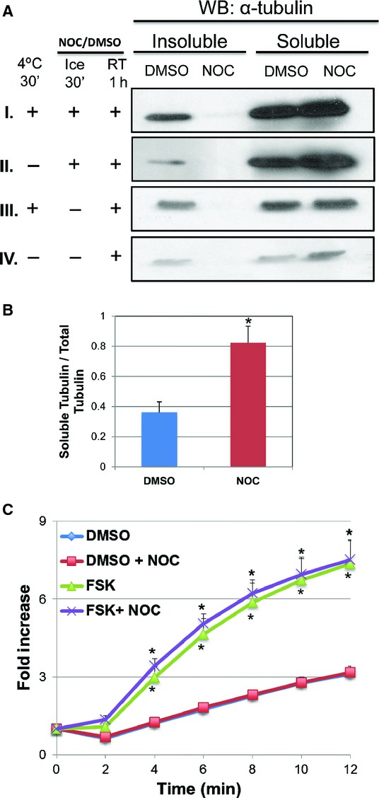
Figure 3.

Effect of microtubule destabilization by nocodazole on forskolin‐induced iodide efflux. (A) HEK‐CFTR cells were pretreated with nocodazole (NOC; 33 μmol/L) for 1 h at room temperature (RT) preceded by either incubation at 4°C for 30′, or addition of NOC for 30′ on ice, or a combination of the two. These representative blots show the 0.1% Triton X‐100 soluble and insoluble α‐tubulin (Ab: monoclonal mouse‐anti‐α‐tubulin; 1:2,500 dilution; estimated size: 50 kDa). (B) The ratio of soluble tubulin to total tubulin (insoluble + soluble tubulin) was analyzed by densitometry of the DMSO and Nocodozole signals for all four conditions. Representative values for protocol ‘I” (the top most blot: 4°C for 30′ → NOC for 30′ on ice →NOC for 1 h at RT) are shown (data not shown for “II to IV”). (C) Effect of nocodazole on iodide efflux in HEK‐CFTR cells. Cells were pretreated with NOC for 1 h at RT during iodide loading, and then stimulated with forskolin (FSK; 10 μmol/L). The data represent the fold change of mean cumulative iodide efflux ± SEM relative to value at starting point. Here n ≥3 for all experiments. DMSO (0.1%) was used as a negative control for NOC and FSK. *P <0.05 versus DMSO control.
