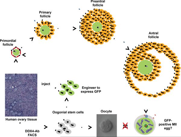
FIGURE 2.
Proposed steps for testing development of fully mature metaphase II (MII) eggs from human OSCs ex vivo. Oogonial stem cells isolated from ovarian cortex differentiate into oocytes in vitro; however, the oocytes formed do not arrest at MII, likely due to a lack of normal interaction with follicular somatic cells. This can be overcome by introducing OSCs into adult human ovarian cortical tissue ex vivo, which leads to the formation of new oocytes contained within primordial follicles (see ref. 16 for more details). After incubation of these micro-thin human ovarian cortical strips in vitro, primordial follicles within the strips activate and progress to primary and preantral stages. Preantral follicles are dissected out and further matured the antral stage, at which time oocytes are isolated for in-vitro maturation to the MII phase (see ref. 60 for more details). To distinguish OSC-derived oocytes from those present in the host tissue, human OSCs are transduced to express GFP prior to injection into the cortical tissue strips. This permits tracking of OSC-derived oocyte formation, primordial follicle assembly and activation, and growth maturation in vitro. If successful, resultant GFP-positive MII eggs derived from human OSCs could then be studied in more detail, including spindle appearance, chromosomal integrity and developmental competency.

