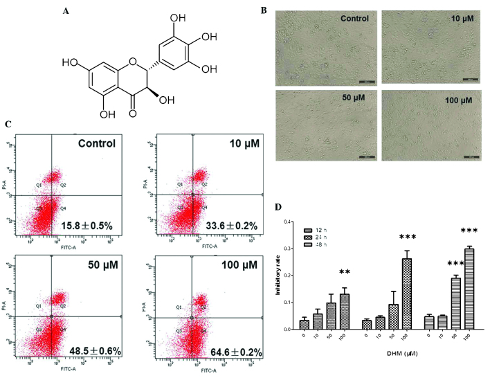
Figure 1.
DHM induces cell growth inhibition and apoptosis in Hepal-6 cells. (A) Chemical structure of DHM. (B) DHM induced cell proliferation in Hepal-6 at various concentrations (10, 50 and 100 μM) for 48 h, visualized by microscopy (magnification, ×100). (C) Hepal-6 cells were treated with various concentrations (10, 50, or 100 μM ) of DHM for 48 h and the results were analyzed by flow cytometry. Each sample was measured in duplicate, and the figure is a representative of three independent assays. (D) MTT assay analyzed cell growth inhibition rates in cells treated with different concentrations (10, 50 and 100 μM) of DHM for 12, 24, 48 h. Values are expressed as the mean ± standard deviation of three independent experiments. **P<0.01, ***P<0.001 vs. 0 μM DHM. DHM, dihydromyricetin; FITC, fluorescein isothiocyanate; PI, propidium iodide; A, area.

