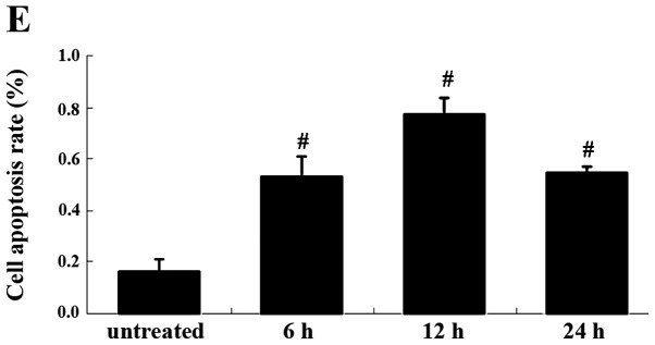
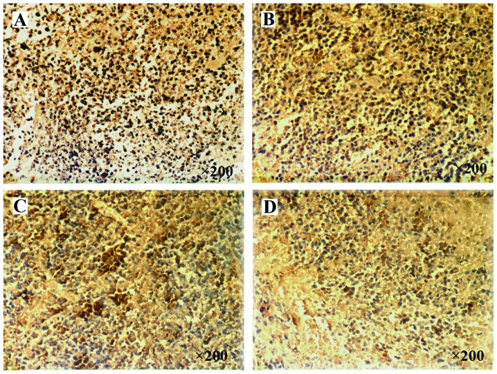
Figure 4.

Apoptotic rate of the cells from human cervical carcinoma in a nude mice xenograft model. A brown-yellow cell nucleus represented an apoptotic cell. (A) Control group; groups at (B) 6 h, (C) 12 h and (D) 24 h, following exposure to picosecond pulsed electric fields (psPEF) treatment. Magnification, ×200 (E) Histogram plotted according to the data in A–D. The data are presented as the means ± standard deviation of three separate experiments. #P<0.01; comparison between two neighboring groups. Following the psPEF treatment, the rate of apoptosis increased, which was most notable in the 12 h group.

