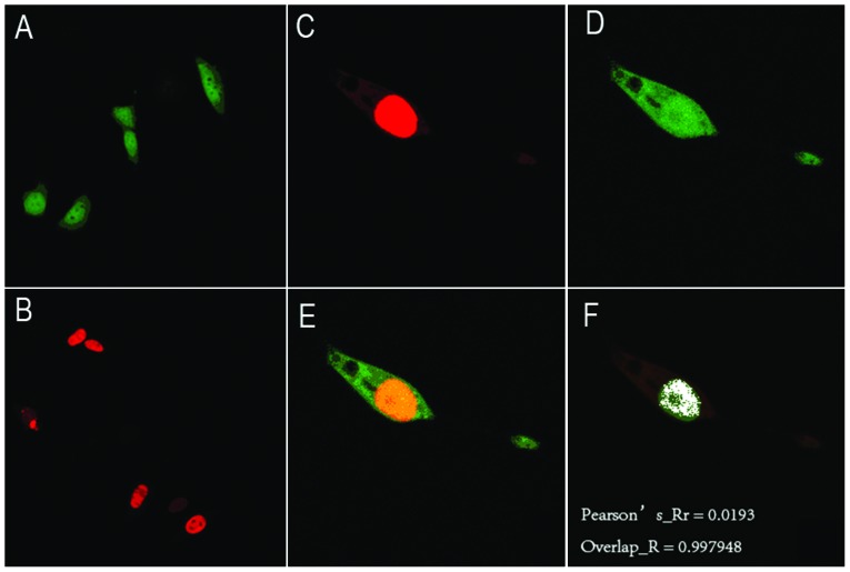
Figure 3.
Colocalization of DND1-β and c-Jun in GC-1 spermatogonia cells by confocal microscopy. (A) GC-1 cells were transfected with pEGFP-c3-Dnd1-β plasmid. Green fluorescence was detected in the nuclei of cells. (B) GC-1 cells were transfected with pmRFP-c-Jun plasmid. Red fluorescence was present in the nuclei of cells. (C–E) GC-1 cells were co-transfected with pEGFP-c3-Dnd1-β and pmRFP-c-Jun plasmids. (C) Red fluorescence was found in the nuclei of cells. (D) Green fluorescence was found in the nuclei of cells. (E) C and D were merged and the overlapped areas of the images are shown in yellow. (F) White areas indicate the co-localization of DND1-β and c-Jun with the following correlation coefficients: Pearson’s Rr=0.0193; overlap R=0.997948; DND, dead end, EGFP, enhanced green fluorescent protein; RFP, red fluorescent protein.

