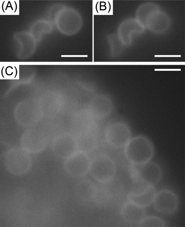
Figure 3.
Fluorescence micrographs of DADM capsules suspended in nematic E7. (A, B) Micrographs of the same group of capsules imaged with different locations of the focal plane such that in (A) the circular capsule on the right of the image was in focus and in (B) the crescent-shaped capsule in the center of the group was in focus. (C) Micrograph of a separate region showing a group of capsules with more spherical than nonspherical capsules. Scale bars are 5 μm.

