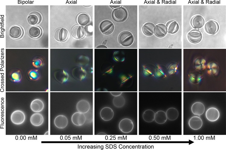
Figure 5.
Bright-field (top row), polarized light (middle row, crossed polarizers), and fluorescence (bottom row) micrographs of representative regions showing LC ordering and wetting in partially filled capsules as a function of increasing SDS concentration (indicated below each column). Labels above each column indicate the internal ordering of the LC at each concentration.

