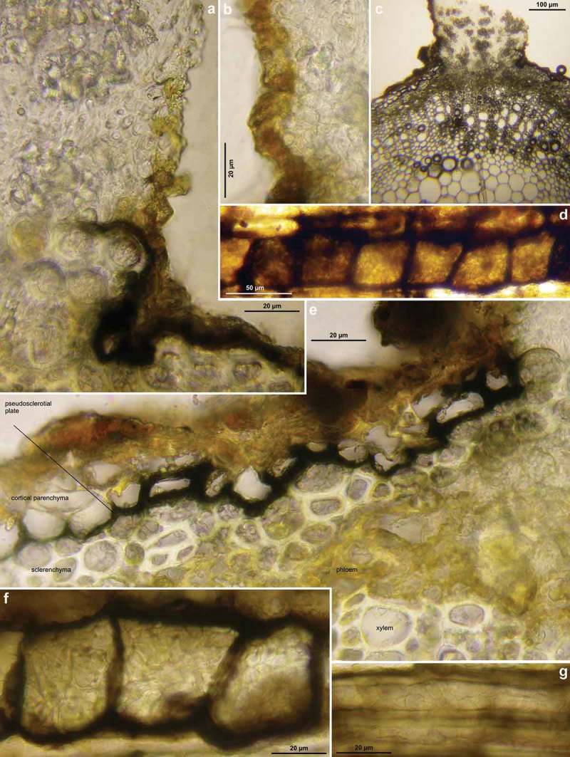
Figure 3.

Hymenoscyphus albidus: a–c, e. median section of stipe base (with internal crystals) and cross section of pseudosclerotium in ash rachis (the black demarcation line is restricted to the border between cortical parenchyma and sclerenchyma, hyphae present in all tissues of petiole); d, f–g. external view on pseudosclerotial plate (tangential section of rachis surface), cells of cortical parenchyma and sclerenchyma densely filled with subhyaline hyphae (textura epidermoidea). – All in living state. – a–g. H.B. 9699 (FR-PC, Granzay-Gript).
