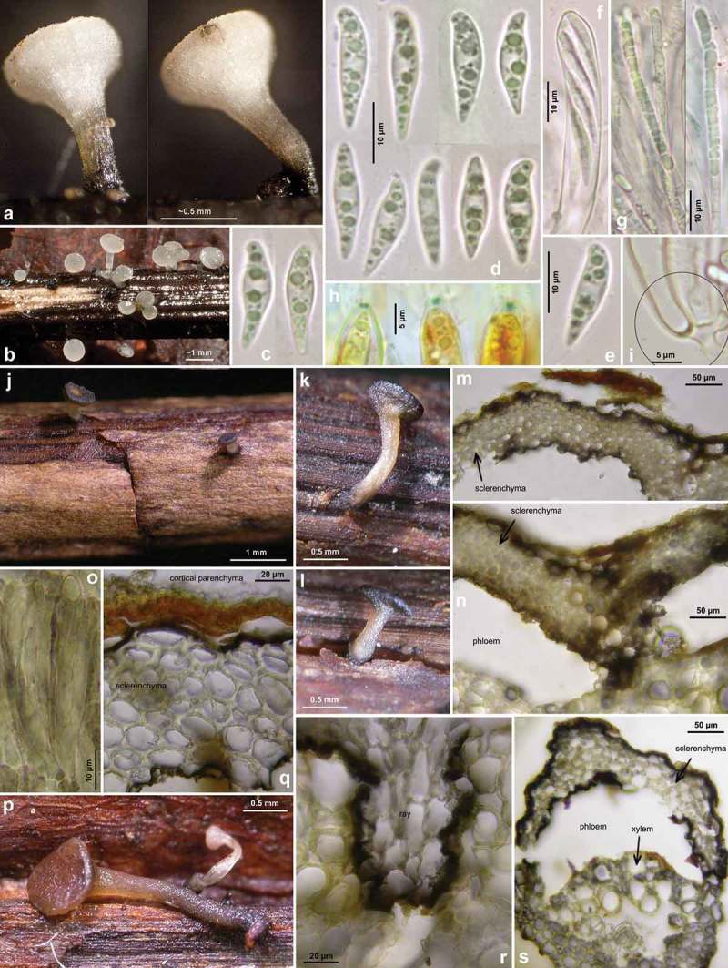
Figure 17.

Hymenoscyphus aesculi: a–b. fresh apothecia; j–l, p. rehydrated apothecia (colour was originally white except for base of stipe); f–g, o. asci and paraphyses(o: with olive secondary pigment especially in paraphyses); h. ascus apices with euamyloid apical ring; i: simple-septate ascus base; c–e. ascospores; m–n, q–s. cross section through cortical region of petioles, with black demarcation line surrounding the sclerenchyma on outer and inner face (cortical parenchyma above and phloem beneath being entirely decomposed). – Living state: c–g; dead state: h (in IKI), o (in H2O). – a–i. E.R.D. 5685 (ES-Asturias, Somiedo); j–o. H.B. 5736 (DE-BW, Gaildorf); p–s. TNS-F-12758 (JP-Honshu, Nagano, Sugadaira). – Phot. a–i: E. Rubio.
