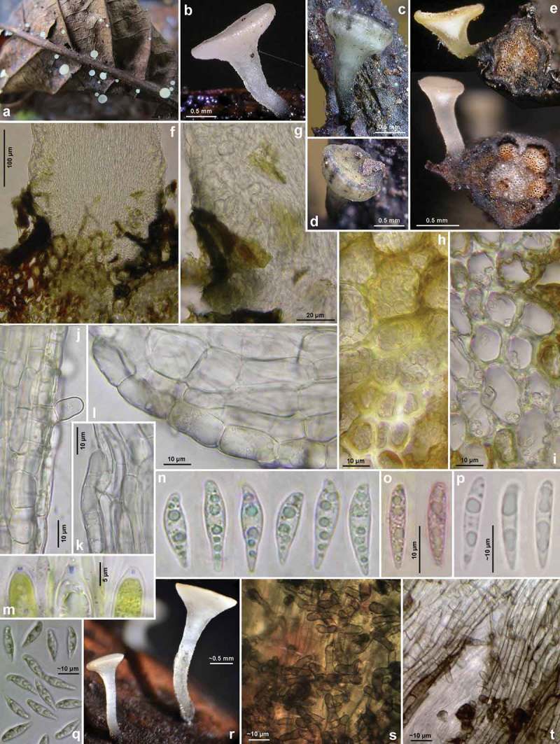
Figure 18.

Hymenoscyphus aesculi: a–b, e, r. fresh apothecia (e: in median section, petioles in cross section); c–d. senescent apothecia (with dark bluish-olive patches on receptacle); f–g. stipe base in median section, showing erumpent growth and absence of crystals; h. cortical parenchyma and sclerenchyma below insertion of apothecial stipe, densely filled with intracellular hyphae; i. sclerenchyma forming a radial ray, loosely filled with hyphae; j–l. ectal excipulum at stipe and (l) at lower flanks in median section, showing VBs in cortical cells; m. ascus apices with euamyloid apical ring; n–q. ascospores (n–o, q: freshly ejected); s–t. surface view on stipe base, showing brown cortical cells and hairs. – Living state, except for m (in KOH+IKI), o (in KOH+CRB), p (in H2O?). – a. 31.VII.2011 (DE-HS, Darmstadt), b–p. H.B. 9701 (Darmstadt), q–t. C.Y. F/2194 (UK-Yorkshire, Halifax). – Phot. a, p: H. Lotz, q–t: C. Yeates.
