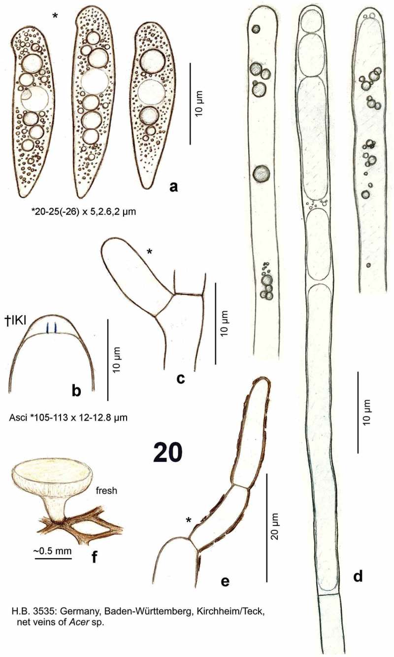
Figure 20.

Hymenoscyphus vacini: a. ascospores (freshly ejected, containing large and small LBs); b. ascus apex with euamyloid apical ring (Hymenoscyphus-type); c. simple-septate ascus base; d. paraphyses containing elongate, low-refractive vacuoles, partly with scattered internal globose VBs of high refractivity; e. cortical hypha of ectal excipulum covered by olive-brown exudate; f. apothecium emerging from net veins of Acer leaf. – Living state (except for b).
