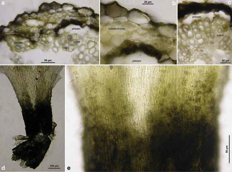
Figure 22.

Hymenoscyphus vacini: a–c. cross section through cortical region of net vein (in H2O), showing black demarcation line surrounding the sclerenchyma on outer and inner face (cortical parenchyma above and phloem beneath being entirely decomposed); fungal hyphae visible inside cells; d–e. apothecial stipe in surface view (slightly squashed, in KOH). – a–c. H.B. 9590 (DE-ST, Merseburg); d–e. H.B. 456 (DE-BW, Stuttgart).
