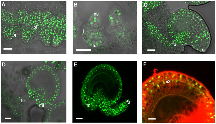
Figure 6. Confocal laser-scanning images of pSTK::STK-GFP expression patterns during ovule and seed development.
(A) Early ovule development: STK-GFP nuclear protein is expressed in the placenta and in the ovule primordia. (B) When integuments arise STK-GFP signal is localized in the nucellus and in the funiculus. (C, D) Mature ovule development: GFP can be detected throughout the integuments, funiculus and the adjacent placental region. (E) After fertilization the STK-GFP signal is present in the outer integuments and funiculus of developing seeds. (F) Magnification of figure E with an overlay projection images of specific PI staining to determine the presence of the GFP signal in the integuments. Protein can be detected in the two layers of the outer integument and also in the more external layer of the inner integument. op, ovule primordia; pl, placenta; nu, nucellus; i, integuments; fu, funiculus; mi, micropyle; ii2, internal layer of inner integument. Scale bars = 50 µm (A and B), 40 µm (C, D and E) and 20 µm (F).

