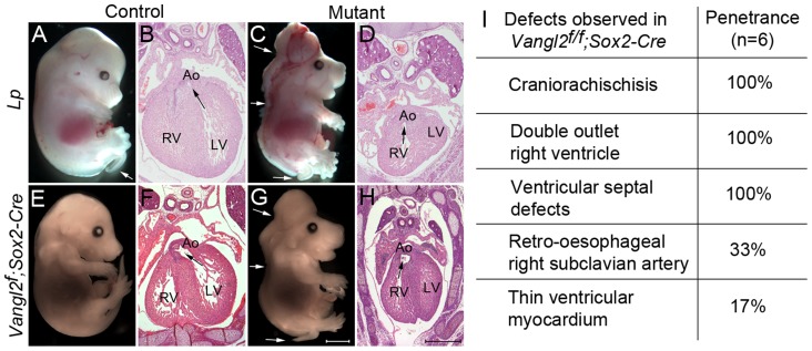
Figure 3. Global loss of Vangl2 recapitulates the Lp/Lp phenotype.
A–D) At E14.5, Lp/Lp embryos exhibit gross abnormalities in body patterning including the severe neural tube defect craniorachischisis (arrows in C). Sectioning of these embryos revealed double outlet right ventricle (the arrows show the communication between the ventricle and the aorta). E–H) The phenotype of the Vangl2flox/flox; Sox2-Cre embryos (G,H) was indistinguishable from Lp/Lp (C,D). The Vangl2f/+; Sox2-Cre however did not have a looped tail, whereas this can be seen in Lp/+ embryos (compare E with A white arrow). I) Breakdown of the cardiac defects seen in the Vangl2flox/flox; Sox2-Cre embryos at E14.5. Also see S3 Fig. Ao – aorta, LV - left ventricle, RV - right ventricle, Vangl2f – Vangl2flox. Scale bar = 2 mm (white), 500 µm (black).

