
Figure 8. Effect of VDCC blockers on membrane potential of unstretched lymphatic muscle.
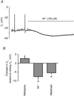
A, original smooth muscle Vm recording in an unstretched rat mesenteric lymphatic vessel illustrating the effect of Ni2+ (100 μm). B, summary bar graph of Vm changes caused by VDCC blockers nifedipine (300 nm), Ni2+ (100 μm) and mibefradil (100 nm). Data are expressed as the calculated differences between Vm in control condition and Vm in the presence of the blocker (n = 4, *P<0.05).