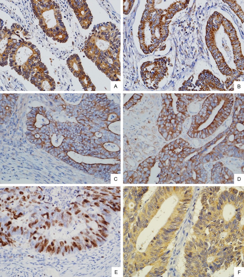
Figure 1.

Immunohistochemical staining of PIK3CA, PIK3CB, MDR-1, LRP, TOPII and GST-π protein expressions in CRC tissues. High-power view (Original magnification ×400) shows strong staining for PIK3CA, PIK3CB, MDR-1, LRP, and GST-π in the cytoplasm of cancer cells (A. PIK3CA; B. PIK3CB; D. LRP; F. GST-π), MDR-1 in the membrane and cytoplasm of cancer cells (C), Topo II in the nucleus of cancer cells (E).
