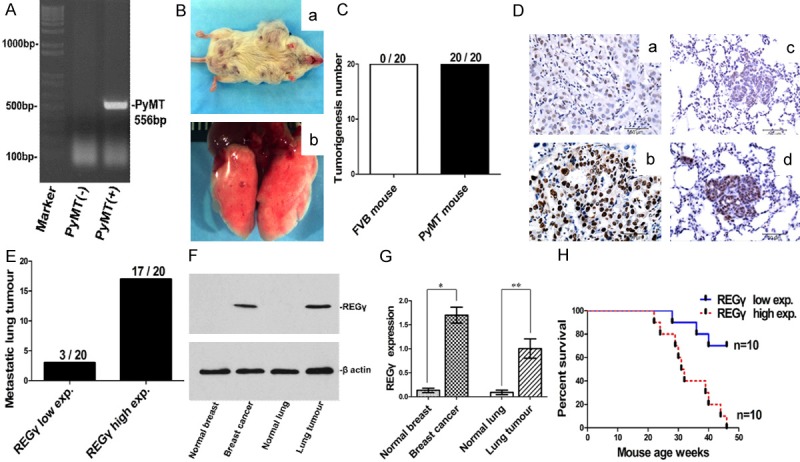
Figure 1.

REGγ expression in mouse mammary carcinoma and metastatic lung tumour. A. DNA genotyping showing MMTV-PyMT transgene band at 556 bp. B. Mammary carcinoma (a) and metastatic lung tumour (b) in MMTV-PyMT mice. C. Tumorigenesis was found in all MMTV-PyMT mice (20/20, 100%) whereas no tumour was found in FVB control mice (0/20, 0%). D. Representative REGγ IHC staining images for mouse mammary cacinoma and metastatic lung tumour (bar = 50 μm). REGγ low expression in mammary carcinoma (a) was associated with low level of REGγ expression in metastatic lung tumour (c), and REGγ high expression in mammary carcinoma (b) was correlated with high level of REGγ expression in the metastatic lung tumour (d). E. Statistical analysis showed that all metastatic lung tumours were REGγ positive (20/20, 100%): 17 metastatic tumours had high level of REGγ expression and 3 tumours had low level of REGγ expression. F. Representative Western blot result of REGγ expression in mouse fresh tissue. G. Densitometry analysis showing relative expression of REGγ as detected by Western blot in fresh samples (20 mice for each group). Statistical analysis showed the significant difference between normal breast tissue and breast cancer tissue (P = 0.0051), as well as between normal lung tissue and metastatic lung tumour (P = 0.0190). H. Survival analysis in a set of MMTV-PyMT mouse (10 mice for each group) showed that mice with high expression of REGγ had significantly poor survival than those with low expression of REGγ (Log-rank Mantel-Cox Test, P = 0.0013, Hazard Ratio = 0.1448, 95% CI of ratio is 0.04474 to 0.4686).
