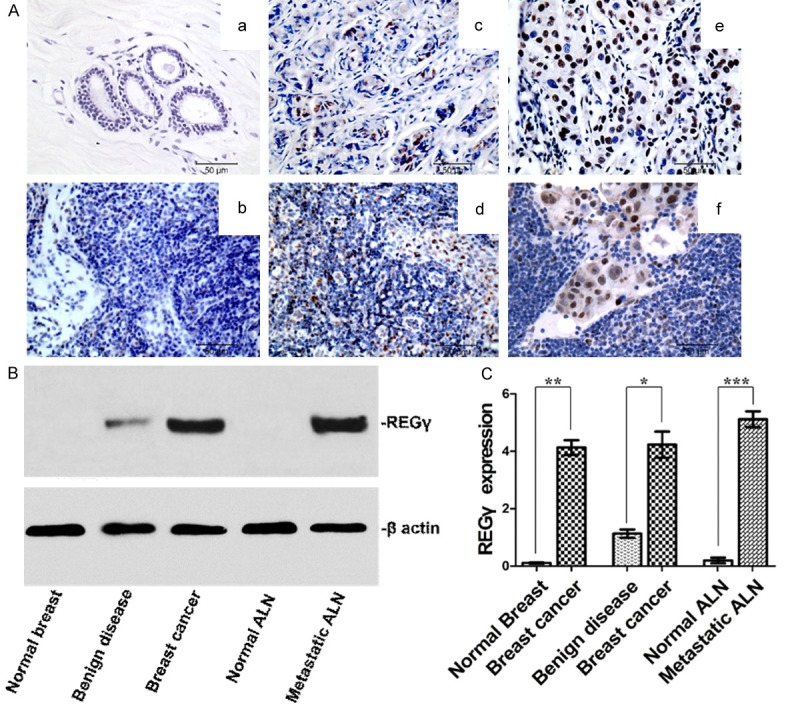
Figure 2.

REGγ expression in human normal breast tissue, normal axillary lymph node (ALN), breast cancer tissue and metastatic ALN. A. Representative REGγ IHC staining images (bar = 50 μm). No REGγ expression was found in normal breast tissue (a) and normal ALN (b). Breast cancer tissue with low expression level of REGγ (c) was associated with low level of REGγ expression in ALN (d). Breast cancer tissue with high expression level of REGγ (e) was correlated with high level of REGγ expression in ALN (f). B. Representative western blot result of REGγ expression in a different set of human fresh tissue. C. Densitometry analysis showing relative expression of REGγ as detected by western blot (30 for each). Statistical analysis showed significant difference between normal breast and breast cancer (P = 0.0012), breast benign disease and breast cancer (P = 0.0024), as well as between normal ALN and metastatic ALN (P = 0.0007).
