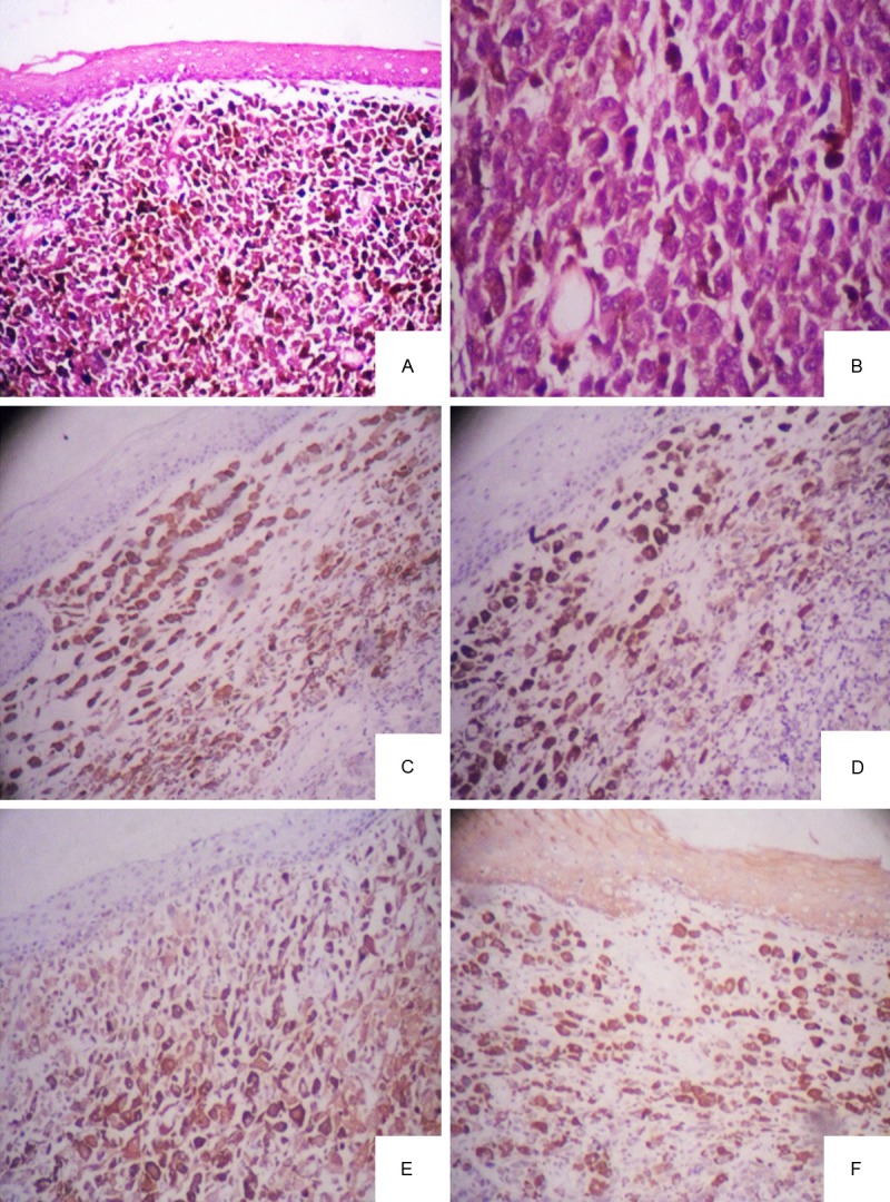
Figure 5.

A. Low power microscopy showed the tumor cells extended downward from the epithelium, melanin deposition (H&E ×40); B. High power microscopy showed tumor cells presented spindle-shapes with abundant cytoplasm, remarkable red nucleoli (H&E ×100); C. Tumor cells were positive to HMB-45; D. Tumor cells were positive to Melan-A; E. Tumor cells were positive to S-100 protein; F. Tumor cells and overlying epithelium were both positive to cytokeratin (CK) pan.
