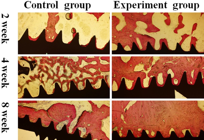
Figure 3.

Histolgical observation and histomorphometric analysis in peri-implant defect by sections of bone tissue combined with dental implant. Original magnification, ×40. In the 2nd week, most of the bone-implant interface without close contact. In the 4th week, most of the bone-implant interface contact directly, mass of newly formed bone can be seen near the interface in experiment group. In control group, reticular formation by new formed trabecular structures within previous bone cavity. In the 8th week, bone tissue was more mature than previous. In experiment group, bone to implant interface was closely contacted, Haversin system could be identified.
